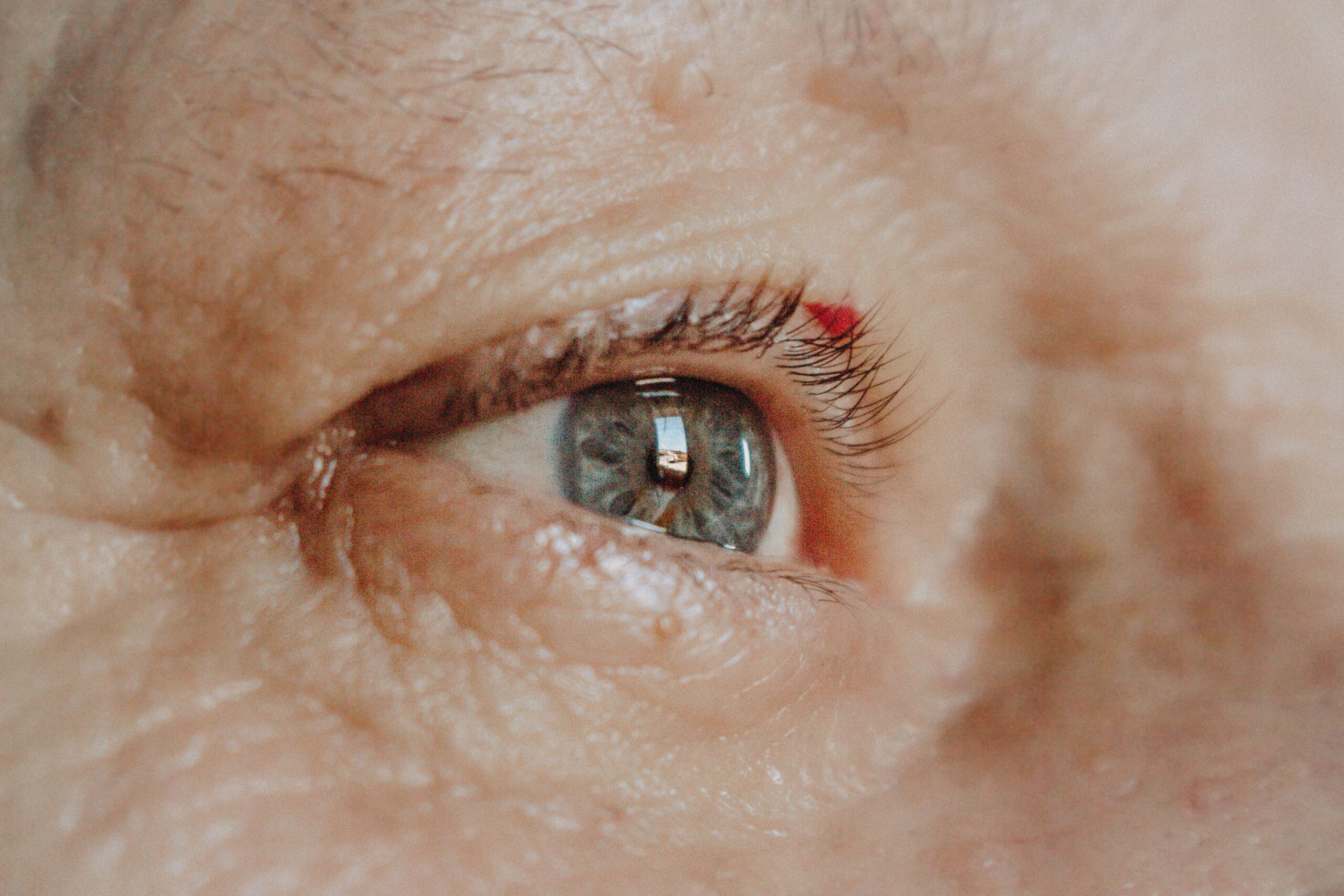

Recognizing your grandchild’s face across the room. Reading without straining toward the light. These are the kinds of moments that depend on your central vision, and they’re exactly what age-related macular degeneration (AMD) can quietly put at risk.
AMD is the leading cause of vision loss in Americans over 50, yet many people don’t know they have it until significant damage has already occurred. Early detection genuinely changes outcomes, which is why understanding the condition and monitoring your eyes between appointments matters as much as the appointments themselves.
What Is Age-Related Macular Degeneration?
AMD is a progressive eye disease that damages the macula, a small but critical area at the center of your retina responsible for sharp, detailed central vision. When the macula is healthy, you can read fine print, recognize faces, and drive safely. When AMD damages it, that detailed central vision deteriorates while peripheral vision typically remains intact.
The result can be disorienting: you might avoid bumping into furniture but be unable to see the person standing in front of you. A road sign is visible ahead, but the words on it aren’t. AMD doesn’t cause complete blindness in most cases, but it can make reading, cooking, watching television, and driving difficult or impossible.

Dry AMD and Wet AMD: Two Very Different Conditions
Dry AMD accounts for approximately 85 to 90 percent of cases and develops slowly over years. Small yellow deposits called drusen accumulate under the retina, gradually damaging the light-sensitive cells in the macula.
The condition moves through recognizable stages. In early dry AMD, a few small drusen are present but most people have no symptoms whatsoever. You wouldn’t know without a comprehensive eye exam. As the disease advances to intermediate dry AMD, more or larger drusen develop, and some patients begin noticing subtle changes: needing more light to read, or slight blurriness in central vision. In advanced dry AMD, also called geographic atrophy, the light-sensitive cells in the macula break down and central vision gradually deteriorates.
Common symptoms of dry AMD include blurred or fuzzy central vision, difficulty reading even with the correct glasses prescription, trouble recognizing faces at a distance, and colors that appear less vivid. What makes dry AMD particularly unpredictable is that it can remain stable for years, then advance, and critically, it can convert to the more aggressive wet form without warning and sometimes without symptoms you’d immediately notice.
Wet AMD affects only 10 to 15 percent of people with AMD, but it accounts for roughly 90 percent of severe vision loss from the disease. In wet AMD, abnormal blood vessels grow underneath the retina in a process called choroidal neovascularization. These vessels are fragile and poorly formed, and they leak fluid and blood into the macula, causing rapid damage to photoreceptor cells.
The warning signs of wet AMD are distinct from dry AMD. Straight lines that suddenly appear wavy or distorted (doorways, window frames, lines of text) are a classic early sign. Patients may also notice a dark or blurry area in the center of vision, rapid changes in clarity, colors that look washed out or different between the two eyes, or difficulty adjusting from bright to low light.
When dry AMD converts to wet AMD, significant vision loss can occur within days to weeks if left untreated. When wet conversion is detected early and treatment starts promptly, vision can often be preserved or even improved. That window is narrow, which is why monitoring between office visits matters so much.
Who's At Risk?
Age is the single greatest risk factor. Risk increases significantly after 50 and continues rising with each decade, with the highest rates in people over 75. Beyond age, family history plays a meaningful role: having a parent or sibling with AMD doubles or triples your risk. Certain gene variants also increase susceptibility, and Caucasians are at higher risk than other racial groups.
Several risk factors are within your control. Smoking is the most significant modifiable risk, with smokers two to three times more likely to develop AMD than non-smokers. Smoking also accelerates progression and reduces treatment effectiveness. Cardiovascular conditions including high blood pressure and high cholesterol increase AMD risk, as does obesity, which roughly doubles the risk of developing advanced AMD. A diet low in antioxidants, omega-3 fatty acids, and dark leafy greens may also contribute, and research suggests that regular exercise has a protective effect.
Treatment Options
There is currently no cure for AMD, but treatment options have advanced considerably, particularly for wet AMD.
For dry AMD, the goal is slowing progression. The AREDS2 formula, developed through the Age-Related Eye Disease Study, has been shown to slow the progression of intermediate to advanced dry AMD. The formulation includes Vitamin C (500 mg), Vitamin E (400 IU), Lutein (10 mg), Zeaxanthin (2 mg), Zinc (80 mg), and Copper (2 mg). These are not standard multivitamins but a precise therapeutic formulation, and they’re appropriate only for patients with intermediate or advanced dry AMD. Talk to your doctor before starting them.
Lifestyle changes also matter. Quitting smoking is the single most impactful step most patients can take. Eating a diet rich in dark leafy greens and omega-3 fatty acids, maintaining a healthy weight, controlling blood pressure and cholesterol, and wearing UV-protective sunglasses all contribute to slowing progression. Research into new treatments for dry AMD, particularly geographic atrophy, is moving quickly, with several therapies currently in clinical trials.
For wet AMD, treatment is available and effective when started quickly. Anti-VEGF injections (vascular endothelial growth factor inhibitors) are the current standard of care. Medications including Lucentis, Eylea, Avastin, Beovu, and Vabysmo are injected directly into the eye using numbing drops, a process that takes only minutes in the office. Most patients describe mild discomfort rather than pain. Injections are typically given monthly initially, then adjusted based on response. When treatment starts early, anti-VEGF therapy preserves vision in the vast majority of patients, and about 30 to 40 percent of patients actually experience improved vision. Delayed treatment significantly reduces that likelihood.
Photodynamic therapy and laser photocoagulation were once more commonly used but have largely been replaced by anti-VEGF therapy for most patients.
The Critical Window: Monitoring Between Visits
The conversion from dry to wet AMD can happen suddenly, and significant vision loss can occur in the weeks between scheduled eye exams. By the time symptoms appear and an appointment is scheduled, permanent damage may already be done.
ForeseeHome is an FDA-cleared home monitoring device designed specifically for this problem. Patients with intermediate dry AMD test their eyes daily, looking into the device for a few minutes per eye while it presents test patterns. The system detects even small changes in central vision distortion that indicate abnormal blood vessel growth, often weeks before those changes would be noticeable to the patient or detectable on a standard Amsler grid. When the device identifies concerning changes, it alerts both the patient and their eye care team so treatment can begin without delay.
The clinical data behind ForeseeHome is compelling. In a major study, 94 percent of patients using ForeseeHome maintained driving vision (20/40 or better) when their dry AMD converted to wet, compared to 62 percent of patients relying on an Amsler grid alone. Detecting wet AMD weeks earlier means starting treatment weeks earlier, and that difference has direct consequences for functional, independent vision.
ForeseeHome is FDA-cleared for patients with intermediate dry AMD in both eyes, intermediate dry AMD in one eye and advanced dry AMD in the other, or advanced dry AMD in one eye with good remaining vision in the other. After an initial training session, the daily test takes three to five minutes and data transmits automatically to a monitoring center. Medicare typically covers it for eligible patients, and many private insurers do as well.
The Amsler grid remains a useful, accessible daily screening tool. Hold the grid at normal reading distance (about 12 to 14 inches), wear your reading glasses if you use them, cover one eye, and focus on the center dot. Take note of whether any lines appear wavy, blurred, or distorted, and whether any areas look blank or dark. Repeat with the other eye, and test daily. Call your doctor immediately if anything changes. The Amsler grid is free and always available, but it has real limitations: by the time changes appear on the grid, some vision damage may have already occurred. It’s a screening tool, not a replacement for ForeseeHome or regular dilated exams.
Living Well with AMD
An AMD diagnosis, particularly one that’s progressing, raises real concerns about independence, driving, and the ability to recognize the people you love. Those concerns are legitimate. So is the fact that most people with AMD maintain enough vision to stay active and engaged, especially with some practical adjustments.
Better lighting makes one of the biggest differences. Increasing light levels throughout your home, particularly in reading and work areas, reduces the strain of compensating for central vision loss. High-contrast colors on stair edges and countertops help with navigation. Organizing your space consistently so items are always where you expect them reduces the cognitive load of reduced vision. Talking clocks, thermostats, and medication reminders are practical tools worth considering.
For reading specifically, magnifying devices range from simple handheld options to electronic video magnifiers that display enlarged text on a screen. Adjusting text size on phones, tablets, and computers costs nothing and helps immediately. Audiobooks and text-to-speech technology have improved significantly and cover most reading needs.
Working with a low vision specialist is worth pursuing earlier rather than later. These professionals teach practical strategies for making the most of remaining vision and can introduce adaptive devices that meaningfully restore independence in daily tasks. Many patients are surprised by what’s possible with the right support.
Vision loss affects emotional health, too. Depression is common among people experiencing significant vision loss, and that’s worth acknowledging directly rather than minimizing. Staying socially connected matters. Support groups for people with AMD exist in many communities and connect patients with others managing the same condition. A counselor experienced with vision loss can provide a different kind of support. Staying as active as possible in hobbies and routines, with whatever adaptations are needed, helps maintain a sense of continuity.
AMD research is moving faster than it was even five years ago. Gene therapy, stem cell approaches, longer-acting anti-VEGF formulations that require fewer injections, and implantable delivery devices are all in active development. Staying connected with your retina specialist is the most reliable way to learn about emerging options as they become available.
What to Do Based on Where You Are
If you’re at high risk but haven’t been diagnosed with AMD, annual dilated eye exams are appropriate starting at age 50, with more frequent monitoring if you have multiple risk factors or a family history of the disease. Addressing modifiable risk factors, particularly smoking, blood pressure, cholesterol, weight, and diet, gives you the best chance of delaying or preventing the condition.
If you have early or intermediate dry AMD, dilated exams every six to twelve months are typically recommended. Daily Amsler grid testing at home is a reasonable baseline, and if you have intermediate dry AMD, ask your doctor whether ForeseeHome is appropriate for you. If your doctor recommends AREDS2 vitamins, start them. Knowing the warning signs of wet AMD conversion in advance means you’ll recognize them if they occur.
If you have advanced dry or wet AMD, close follow-up with your retina specialist is essential. Missing anti-VEGF injection appointments carries real risk of vision loss, so keeping that schedule is a priority. Report any vision changes promptly, even ones that seem minor. Working with a low vision specialist helps you make the most of remaining vision while treatment continues.
Regardless of where you are in the disease, certain symptoms require same-day contact with your Charlotte eye doctor: sudden appearance of wavy or distorted straight lines, new dark or blank spots in central vision, a rapid drop in clarity, or a sudden increase in floaters or flashes. Don’t wait to see whether these resolve on their own. With wet AMD, the hours and days before treatment begins matter.
Your Partners in Preserving Vision
Our retina specialists diagnose and manage both dry and wet AMD using advanced diagnostic technology and current treatment protocols. We partner with ForeseeHome to offer intermediate dry AMD patients home monitoring between visits, and Horizon Eye Care welcomes patients seeking a second opinion or transitioning care from another practice.
To schedule a consultation with one of our retina specialists, call (704) 365-0555. With seven locations across the Charlotte area, we make every effort to see patients quickly when vision-threatening changes occur.